
Simulation-based Ultrasound Training Supported by Annotations, Haptics and Linked Multimodal Views
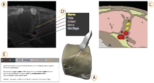
When learning ultrasound (US) imaging, trainees must learn how to recognize structures, interpret textures and shapes, and simultaneously register the 2D ultrasound images to their 3D anatomical mental models. Alleviating the cognitive load imposed by these tasks should free the cognitive resources and thereby improve the learning process. We argue that the amount of cognitive load that is required to mentally rotate the models to match the images to them is too large and therefore negatively impacts the learning process. We present a 3D visualization tool that allows the user to naturally move a 2D slice and navigate around a 3D anatomical model. The slice is displayed in-place to facilitate the registration of the 2D slice in its 3D context. Two duplicates are also shown externally to the model; the first is a simple rendered image showing the outlines of the structures and the second is a simulated ultrasound image. Haptic cues are also provided to the users to help them maneuver around the 3D model in the virtual space. With the additional display of annotations and information of the most important structures, the tool is expected to complement the available didactic material used in the training of ultrasound procedures.
